The Neuroscience of Seizures

What are Grand Mal Seizures?

Imagine first aid responders, dressed in red, running across the field in urgency on a typical day at summer camp. These are the series of events that I witnessed during my first week as a counsellor, hanging out with my cabin on that same field. Seeing all the commotion, I asked another counsellor, “what is going on?”

They responded, “a camper is having a seizure”.

Immediately, my mind went to the stereotypical images of what a seizure looks like: Jerking movements, eyes moving from side to side and muscles twitching.

I went home that night, opened my computer and fell into a rabbit hole of research on “what is a seizure?”

The first statistic that popped up on my screen was “about 1 in 10 people will have a seizure in their lifetime”. I was shocked to read this statistic and thought to myself, “IT’S THAT COMMON!?”

I later learned that there are many types of seizures. Some may present as blank stares into space, while others may present as the stereotypical seizures often seen on TV and that I thought of when I heard about the camper.

Grand mal seizures, also known as tonic-clonic seizures, are the most recognizable type of seizure. They account for 25% of all generalized seizures and often occur with epilepsy.

In this blog, I am going to write about how to recognize a tonic-clonic seizure, its causes, what happens in the brain and what you can do to help.

How to Recognize a Tonic-Clonic Seizure

Tonic-clonic seizures are a type of generalized seizure that results in a loss of consciousness and muscle contractions.

Tonic-clonic seizures may begin with a simple partial seizure, where the person may experience changes in sensation and mood. The partial seizure may then spread from one side of the brain to the other, resulting in a tonic-clonic seizure.

During the tonic phase:

  • Muscles stiffen and become rigid

  • The person may let out a cry or groan as the muscles in the body contract, forcing air out of the vocal cords.

  • Loss of consciousness

During the clonic phase:

  • The arms and legs begin to jerk rhythmically

  • Breathing may be limited

  • Blue lips or a dusky-looking face

  • The jerking movements will slow before the seizure stops and consciousness will gradually return.

After tonic-clonic seizures, the individual may experience the following symptoms:

  • Confusion

  • Drowsiness

  • Irritability

  • Loss of memory about the seizure episode

  • Headache

  • Todd paralysis (weakness on one side of the body, lasting a few minutes to a few hours)

How to Respond When You See Someone Having a Tonic-Clonic Seizure?

When I heard a camper was having a seizure, I was in shock. I thought to myself, what would I do if that was one of my own campers and truthfully, I did not know how to answer that question. I now understand how important it is to know how to respond when you encounter someone having a tonic-clonic seizure, as it could potentially save their life.

Do not touch or try to restrain the person when they are having a seizure.

Do not put anything in the individual’s mouth.

Do move objects away from the individual to minimize the risk of them injuring themselves.

Do time the seizures. Generally, tonic-clonic seizures last 1 to 3 minutes; however, if they last more than 5 minutes, or three seizures occur in a row, immediately call 911 for emergency help.

Do provide support to the individual once the seizure stops as they may feel confused or tired.

Causes

Tonic-clonic seizures can be caused by many conditions, such as:

  • Epilepsy

  • Traumatic brain injury: Some people develop a seizure after a traumatic head injury

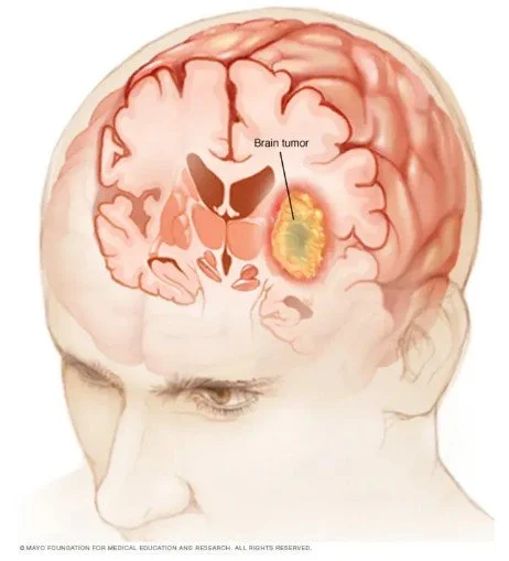
  • Brain conditions: Strokes and brain tumors may result in brain damage, causing seizures

  • Congenital or developmental factors: Genetic syndromes or blood vessels that do not fully develop within the brain can lead to a seizure

  • Metabolic problems: Low levels of blood glucose, sodium, calcium or magnesium may increase the likeness of having a seizure

  • Extreme lack of sleep

For a more extensive list on the causes of seizures, visit this link: https://www.epilepsy.com/causes

What Happens in the Brain?

Neurons within the brain communicate by transmitting electrical impulses from one neuron to another. In a healthy brain, neurons fire independently and at different times. Seizures occur when there is an abnormal release of electrical activity by a large group of neurons firing at the same time. In tonic-clonic seizures, both hemispheres of the brain are affected. The abnormal release of electrical activity interrupts normal brain function. The subcortical, cortical and brain stem networks are affected.

For more information on what happens in the brain during a seizure, watch this video: What Happens in Your Brain During a Seizure | WebMD

Diagnosis of Epilepsy

An electroencephalogram (EEG) is used to detect abnormalities in brain waves or electrical activity of the brain. EEG’s can be used to detect seizure activity in the brain as it will appear as spike-and-wave discharges on the EEG. The spike-and-wave discharges on the EEG result from synchronized bursts of neuronal firing that occur during a seizure. However, the sensitivity for an EEG to identify a seizure is less than 50% and therefore, a normal EEG does not rule out the possibility that the patient did not have a seizure.

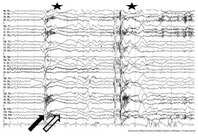
Figure 1: EEG of a Tonic Seizure.

I hope you found this blog informative. Shortly, Jaiden will be posting another blog where he will dive deeper into the neuroscience and treatments for seizures. Stay tuned!

-Jordy

References

  1. https://www.cdc.gov/epilepsy/about/first-aid.htm#:~:text=About%201%20out%20of%2010,the%20seizure%20stops%20by%20itself.

  2. https://my.clevelandclinic.org/health/diseases/22788-tonic-clonic-grand-mal-seizure

  3. https://www.epilepsy.com/diagnosis/eeg/what-if-its-normal#:~:text=A%20normal%20EEG%20does%20not,have%20a%20normal%20EEG%20test.

  4. https://www.ncbi.nlm.nih.gov/books/NBK390347/figure/f71/

  5. https://www.epilepsy.com/diagnosis/eeg#:~:text=EEG%20is%20an%20important%20test,wires%20to%20an%20electrical%20box.

  6. https://www.sailforepilepsy.org/voices-for-epilepsy/seizure-first-aid/?gad_source=1&gclid=CjwKCAiA04arBhAkEiwAuNOsIu6_MRfPygidWs1bj-40bGFxFjA-D1Z3NB3vKkFhtBAgM6sfCMJz9hoCdwgQAvD_BwE

  7. https://www.uchicagomedicine.org/conditions-services/neurology-neurosurgery/epilepsy-seizures/causes#:~:text=Experts%20believe%20that%2C%20in%20many,fold%20increased%20risk%20for%20epilepsy.

  8. https://www.hopkinsmedicine.org/health/conditions-and-diseases/epilepsy/tonic-clonic-grand-mal-seizures#:~:text=Tonic%2Dclonic%20seizures%2C%20formerly%20known,phase%20and%20a%20clonic%20phase.

  9. https://www.epilepsy.com/what-is-epilepsy/seizure-types/tonic-clonic-seizures

  10. https://www.cedars-sinai.org/health-library/diseases-and-conditions/t/tonic-clonic-grand-mal-seizure.html

  11. https://www.ncbi.nlm.nih.gov/books/NBK554496/#:~:text=A%20generalized%20tonic%2Dclonic%20seizure%20is%20a%20motor%20seizure%20and,brainstem%20networks%20of%20the%20brain.

Transcranial Ultrasound Stimulation and its Applications in Treating Brain Tumors

Introduction

What if we could ablate a brain tumor without needing to cut into the brain? There are currently many techniques being developed that would non-invasively treat brain tumors. One of these technologies is a high-intensity focused ultrasound that has recently been approved by the FDA for the treatment of essential tumors.

There are more than 120 different types of brain tumors that differ in the type of cells that they are made of and where they occur. Brain tumors may be benign (noncancerous) or malignant (cancerous). Treatment options for brain tumors depend on the individual’s symptoms, preferences, along with the type, size and location of the tumor.

Some examples of treatment options include surgery, radiation therapy, chemotherapy and drug therapy. Multiple factors, such as the brain’s natural defenses and the blood-brain barrier (BBB) pose a major hurdle for the treatment of brain tumors. As well, if the tumor is near a delicate part of the brain, surgery may be difficult. Transcranial ultrasound stimulation (TUS) is an emerging technique and approach to non-invasively treat brain tumors. TUS can be used with converging methods of treating brain tumors.

Figure 1: Brain Tumor — A growth of cells that occur near or within brain tissue.

What is TUS?

Transcranial ultrasound stimulation (TUS) is an emerging technique that is non-invasive and has good spatial resolution. TUS is a hemispherical phased-array transducer that produces ultrasonic frequency. Ultrasonic frequency is anything above human hearing, which is about 20,000 hertz.

TUS has many different applications within neuroscience. At low intensity (< 100 W/cm2), TUS delivers short bursts of sonic waves to affect the voltage-dependent neural circuits in the brain. The tool can modulate neural circuits by inhibiting or exciting neural activity. The process of neuromodulation can assist in modulating symptoms of neurodegenerative diseases, strokes or epilepsy. Medium intensity (100–200 W/cm2) ultrasound has been used to increase the opening of the blood-brain barrier. High intensity (> 200 W/cm2) focused ultrasound has the ability to induce permanent lesions and treat some essential tumors.

How does TUS work to treat brain tumors?

There has been an increasing amount of research in how transcranial ultrasound stimulation can be used to treat brain tumors or increase the efficacy of other treatment options.

By focusing ultrasound waves at a single point and at a high intensity, TUS can ablate tumors or cause permanent lesions. This can occur through thermal ablation or the coagulation of cellular proteins. Thermal ablation occurs by projecting an ultrasound beam into the brain, resulting in elevated tissue temperatures within the focal zone or around the tumor.

Transcranial ultrasound stimulation can also be used to assist in drug delivery. The blood-brain barrier (BBB) is a layer of blood vessels that filter the blood flowing into the brain. The barrier makes it harder for some substances, such as anticancer drugs to enter the brain. BBB is a major obstacle limiting the therapeutic efficacy of drugs to treat brain tumors. TUS at a medium focused intensity can increase opening of the blood-brain barrier for drug delivery. One study has shown that TUS can enhance CNS drug delivery by opening the BBB in animal models, along with enhancing both innate and adaptive immunities. Strong innate and adaptive immunities are essential to orchestrate an antitumor response.

With the ability for TUS to thermally ablate brain tumors and increase the efficacy of other treatment options, TUS acts as a promising technique for patients. NaviFUS is a new TUS device that transcranially delivers ultrasonic energy into deep tissues of the brain, while being guided by a neuro navigator. The device is mobile, not limited to the operation room, has a short procedure time (<30 min) and is cost effective. NaviFUS has been approved by the FDA .

What is the procedure of NaviFUS?

NaviFUS: Introduction

I hope you found this blog informative. Remember that treatments for brain tumors differ based on their characteristics. Always consult with a doctor about the most appropriate treatment options.

-Jordy

References

https://brainbox-neuro.com/techniques/tus

https://www.fusfoundation.org/diseases-and-conditions/brain-tumors-glioma-and-metastatic/

https://navifus.com/about/

https://pmc.ncbi.nlm.nih.gov/articles/PMC7080303/

https://www.mayoclinic.org/diseases-conditions/brain-tumor/symptoms-causes/syc-20350084

Deciding When

Imagine the hypothetical scenario in which you are forced to choose one food to eat for the rest of your life, with the choices being between a Twix candy bar or Coffee Crisp. This may seem like a small, simple choice, but this is the only thing you will be allowed to eat for the rest of your life, so you have to put some thought into this decision. This decision will be based on memories you have associated with each option, whether you enjoyed the taste of Twix, or Coffee Crisp made you throw up one time when you were younger.

The underlying processes in decision making are fundamentally the same in all types of decisions, from simple choices like choosing to study at Stauffer Library or Douglas Library for the day, to life changing decisions such as the example of the chocolate bars. The brain is presented with a decision, so it will retrieve past memories to base it off of. If for example, you have been very productive at Douglas Library, rather than at Stauffer, then your prefrontal cortex will make this distinction and choose to spend the rest of the day at Douglas.

Prefrontal Cortex (blue) relative to the rest of the brain. Source: Maher, Courtney. “Prefrontal Cortex Damage: What to Expect & How to Recover.” Flint Rehab, 18 Aug. 2020, www.flintrehab.com/prefrontal-cortex-damage/.

The prefrontal cortex is the main structure involved in these processes, with its primary function to store and retrieve short term memories, although it works closely with the hippocampus to use these memories.

Hippocampus (blue) relative to the rest of the brain. Source: Rehab, Flint . “Hippocampus Damage: Effects, Treatment & Recovery.” Flint Rehab, 1 Apr. 2020, www.flintrehab.com/hippocampus-brain-injury/.

There are many other regions in the brain involved during thought processes, although the identification of the exact functions remains a large gap in neurophysiology.

These processes occur in four main steps, where we start with an initial sensory stimuli that transmits a signal to the brain via sensory neurons. Sensory neurons receive information from the outside world and convert it into action potentials for the brain to interpret. Going back to the chocolate bar example, when you are told that you must choose between both chocolate bars, sensory neurons take in this information and process it to the brain. A second stimulus further excites the hippocampal neurons, causing an initial neural response from the collection of both stimuli, which goes on to activate the prefrontal cortex. This stimulus comes from other information necessary to make the decision. So if it is a person telling you you have to choose between Twix and Coffee Crisp, these secondary stimuli will take in the speaker’s tone, body language and use of words to evaluate the situation. The PFC then retrieves relevant past information to make the decision and relate them to the situation. If you had an allergic reaction last time you had a Coffee Crisp, then this memory will get brought into the equation. In the final step, the PFC commits to the necessary course of action for the decision made. This is mediated by motor neurons that send the signal from the brain to the muscles, to pursue the intended path. Finally, after much thought, your brain chooses Twix over Coffee Crisp, because as we all know, Twix is much better than Coffee Crisp, hence the motor neurons will send signals to your muscles, to indicate that you chose Twix.

You might be asking yourself, well how fast does this process take place, how much time does it take for us to receive a signal and carry through our chosen course of action? When we are driving for example, we need these decisions to be made fairly quickly, or else we would all end up in the hospital. Benjamin Libet, a prominent neuroscientist in human consciousness research, studied the timing as to when do we actually make the decisions and carry out their respective actions. He claims that the decisions we make are predetermined, which means that our brain has already made the decision before we become consciously aware of the decision we make. Benjamin Libet’s contributions to decision making processes can be adapted to the philosophical debate on free will. He showed scientific evidence supporting determinism, but there is much controversy in the accuracy of the data. So if our biology knows our every move, does it disprove free will? But this is a topic for another time…

Feel free to write back if you have any comments, questions, concerns or any ideas as to what you want us to write about.

Jaiden Plante

The Hunt for a Cure: An Overview of the Medication of Seizures

The uncontrollable jerking of limbs, loss of consciousness, and lifeless stares that accompany seizures have been well known for millennia, but it was only until recent scientific past, that humans have been able to describe the biochemical background behind them. From a neuroscientific perspective, seizures can be described as involuntary neuronal firing, occurring either in a localized area, or throughout the entire brain. These irregular electrical impulses in the brain stimulate the abnormal physical and emotional behavior experienced by seizure patients, meaning the symptoms have a dependence on the locality at which this unwanted firing of neurons is taking place. Any scientist working in this field would ask the obvious question of “why does this happen”, and there are many reasonable answers, which include brain tumors, strokes, and chemical imbalances. Unfortunately, there are many cases in which there may be no cause, and when these types of seizures occur multiple times in a short period of time, they are characterized as an epileptic disorder. This unresolved causation for epilepsy has led epileptic researchers to focus on lessening the symptoms for the patients, which is done primarily through the use of drugs.

Antiepileptic drugs (AED’s) have been used effectively for over a century, but there will always be ways to improve them and lessen the symptoms for the patients. With the thousands of drugs that have been developed, they can be classified based on their mechanism of action.

The most common AEDs are grouped as sodium channel blockers, and they do exactly as their name suggests. They are part of the anticonvulsant class of medication, which suppresses neuronal activity. To understand what this involves, the basics of neuron activation, or more commonly known as action potentials, must be acknowledged. Action potentials, the firing of a neuron in the brain, are mediated by sodium channels, found on the cell membrane. These channels open and close allowing the diffusion of sodium cations (positively charged ions), which transmit the signal across the neuron. As the positive sodium ions enter the cell, they create a rapid change in voltage, which induces an electrical charge within the cell. This electrical current can then be passed on to other neurons and transmit a signal from one part of the body to another. In AED’s, sodium channel blockers inhibit the activation of sodium channels, which prevents the sodium ions from entering the cell, which maintains the resting state voltage of the cell, and disallows for a signal to be emitted. This can be very beneficial during seizures, when there is involuntary neuron activity. Some of the main sodium channel blockers include:

  • Carbamazepine

  • Phenytoin

  • Fosphenytoin

  • Oxcarbazepine

  • Eslicarbazepine

  • Zonisamide

For more information of the science behind sodium channel blockers and how they help suppress epileptic symptoms, feel free to read Voltage gated sodium channel inhibitors as anticonvulsant drugs.

Another common group of AED’s used in epileptic treatment are GABA reuptake inhibitors, and once again, they do exactly as the name suggests. The basics of neuron communication must be understood to fully grasp the concept of GABA reuptake inhibitors. When a neuron is activated, it will transmit its signal to other neurons by sending neurotransmitters to the target cell, which is known as the postsynaptic neuron, through a synaptic cleft. These neurotransmitters will bind to receptors on the postsynaptic cleft and transmit the signal to the next cells. Once neurotransmitters have fulfilled their role, they will be brought back into the original cell through a process known as reuptake. GABA, short for gamma-aminobutyric acid, is an inhibitory neurotransmitter that suppresses the activity of neurons. These neurotransmitters will bind to chloride channels, sending in negatively charged chloride ions into the cell. This process known as inhibitory postsynaptic potential (IPSP’s), lowers the probability of the neuron becoming active, since it lowers the voltage of the cell even further. Normally, GABA will be brought back by the pre-synaptic cell, so it can be recycled. With GABA reuptake inhibitors, this process is terminated, and the GABA neurotransmitters are left in the synaptic cleft to bind to more receptors and further hyperpolarize the cell. In epilepsy, this lowers the chance neurons are activated at random, reducing epileptic symptoms. One of the main GABA reuptake inhibitors is Tiagabine and it is an anti-convulsant that can also be used as anxiety medication.

For more information on the chemical basis of GABA reuptake inhibitors feel free to read GABA Reuptake Inhibitor.

These are only two types of the many drugs used deal with epilepsy, while many other types of AED’s deal with the same underlying principles associated with sodium channel blockers, and GABA reuptake inhibitors. The variety in AED’s allows physicians to accommodate for each individual and their needs to find the right prognosis, since there is no one drug that will completely cure epilepsy. Other treatments for epilepsy deal with the surgical removal of the area of the brain that seizes, which is known as resective brain surgery, or deep brain stimulation which involves putting a device in the body, that sends signals disrupting the electrical impulses emitted by the seizure. There will always be better ways to deal with this disorder, thanks to the advancements in technology, so we should keep our hopes up and not give up on finding a cure for epilepsy.

If you have any questions, concerns, comments, or ideas on epilepsy, seizure and medications, or just Neuroscience in general, feel free to write back to us. We would love to hear your ideas and opinions to help educate and inform the community about the beauty of neuroscience.

-Jaiden